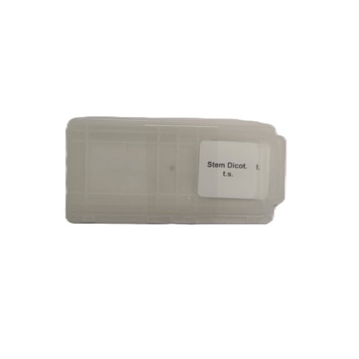
Microscope Slide - Stem Dicot. t.s. Individually packed in PP slide mailer
SL-OPT-029
Vendor: SmartLabs
Couldn't load pickup availability
- Fast Nationwide Delivery
- 100% Quality Guaranteed
- Reliable & Secure Payments
Have a question about this product?
Send us a message and we'll get back to you.
This prepared microscope slide of dicot stem (T.S.) comes individually packed in a PP slide mailer. It's made using professional biological sectioning techniques, with every step carefully controlled, from collecting and fixing the sample to dehydration, embedding, sectioning, staining and mounting.
The slide features a transverse section (t.s.) of a dicot stem, allowing observation of its internal structure. The section typically shows key anatomical features such as the epidermis, cortex, vascular bundles arranged in a ring, and pith, illustrating the organisation characteristic of dicotyledonous stems.
Use in Education
Ideal for botany and general biology courses, this slide supports lessons on plant anatomy, stem structure, and the arrangement of vascular tissues. It can be used in classroom demonstrations and laboratory exercises to help students compare monocot and dicot stems. Students can also practise microscope focusing and observation techniques while exploring tissue organisation in dicot stems.
How the Slides Are Made
- Raw material: Only fresh, undamaged biological tissue is selected, so the sections are authentic and scientifically accurate.
- Preservation: Professional fixatives preserve the samples quickly, so the tissue cells don't deform or deteriorate. The tissue then goes through graded dehydration, clearing and wax infiltration, which gives it an even texture and the right hardness for sectioning. Each step is carefully timed and uses controlled reagent concentrations.
- Sectioning: High-precision sectioning machines, adjusted by experienced technicians, give an even section thickness with no wrinkles or damage. Sections are usually 5 to 10µm thick.
- Staining and mounting: Classic methods such as haematoxylin and eosin (H&E) staining, carefully timed, give clear contrast between nucleus and cytoplasm and clear colours. A high-transparency resin bonds the section to the slide and cover glass, keeping out moisture and oxidation, so the slide can be stored and reused for a long time without the colours fading or the section lifting.
- Please note that the steps may vary depending on the specimen.
Useful Slide Abbreviations
- T.S. or C.S. = transverse or cross section: a thin slice across the organism or tissue.
- L.S. = longitudinal section: a slice along the length of the organism.
- C.S. & L.S. = cross and longitudinal sections on the same slide for comparison.
- W.M. = whole mount: the entire organism or structure is mounted on the slide.
Product Specifications
- Material: Glass slide with specimen, neutral gum resin and a glass cover slip.
- Slide: Microscope slide 7101 (non-frosted), ground edges, 45° corners.
- Cover glass: Round, 16mm.
- Sectioning: Usually prepared on a sectioning machine, depending on the specimen.
- Resin: Neutral gum.
- Packaging: Individually packed in a PP slide mailer.
- Please note specifications may vary.