Model Animal Cell Division Meiosis - Mini
ZM16083
Vendor: EISCO
Couldn't load pickup availability
- Fast Nationwide Delivery
- 100% Quality Guaranteed
- Reliable & Secure Payments
Have a question about this product?
Send us a message and we'll get back to you.
This set of 12 models illustrates the detailed stages of meiosis in an animal cell, designed to show the cellular structures as seen under a microscope. These stages ensure that each resulting haploid daughter cell receives a unique combination of chromosomes from the parent cell. The models are coloured to distinguish the different cellular components and are mounted on a base for classroom demonstrations and individual study.
Size 42 x 30 x 5 cm approx. Weight 940 g approx.
Prophase I
Chromosomes condense, homologous chromosomes pair up (synapsis), and crossing over (exchange of genetic material) occurs. Prophase I is further divided into five substages:
- Leptotene: the first stage, characterized by the condensation of chromosomes into thread-like structures within the nucleus. Homologous chromosomes begin to pair up, although they are not yet fully synapsed (associated).
- Zygotene: homologous chromosomes (one from mother and one from father) come together and pair. This pairing is known as synapsis, and it is very specific and exact.
- Pachytene: homologous chromosomes are fully paired and synapsed, meaning they are tightly connected along their lengths by the synaptonemal complex. This stage is also characterized by crossing over, where genetic material is exchanged between non-sister chromatids of homologous chromosomes.
- Diplotene: the fourth stage, during which the paired chromosomes begin to separate into two pairs of chromatids.
- Diakinesis: the fifth and last stage, when the separation of homologous chromosomes is complete and crossing over has occurred. The chromosomes are in their most robust state, and in a bivalent there is still at least one chiasm connecting the homologous chromosomes. The centrosome moves to the equator, the nucleolus dissolves, the nuclear envelope collapses, and the mitotic spindles move.
Rest of meiosis I
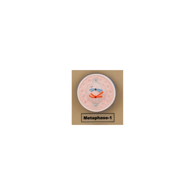
- Metaphase I: homologous chromosome pairs line up at the cell's equator (metaphase plate).
- Anaphase I: homologous chromosomes are separated and pulled to opposite poles of the cell.
- Telophase I: chromosomes arrive at the poles, nuclear membranes may reform, and the cell divides (cytokinesis I).
Meiosis II
- Prophase II: chromosomes condense again if they decondensed in Telophase I, nuclear envelopes break down, and new spindle fibers form.
- Prometaphase II: spindle fibers attach to the chromosomes.
- Metaphase II: chromosomes line up individually at the equator of each cell.
- Anaphase II: sister chromatids of each chromosome are separated and pulled to opposite poles.
- Telophase II: chromosomes arrive at the poles, nuclear envelopes reform, and the cell divides (cytokinesis II).